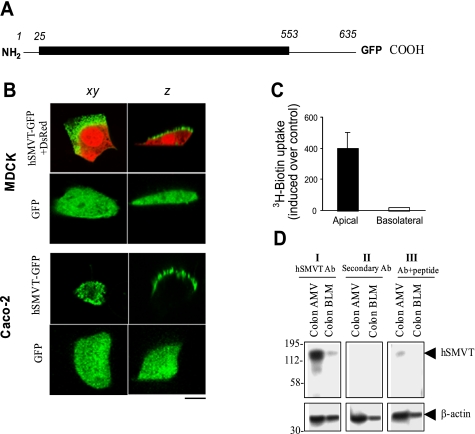
Fig. 1.
Apical targeting of the human sodium-dependent multivitamin transporter (hSMVT)-green fluorescent protein (GFP) fusion protein in renal and intestinal epithelial cells. A: schematic representation of the full-length hSMVT protein (1-635 residues) with GFP fused to the COOH terminus (hSMVT-GFP). B: distribution of hSMVT-GFP and GFP alone in lateral (xy, left) and axial (z, right) section in renal [top; Madin-Darby canine kidney (MDCK)] and intestinal [bottom; human adenocarcinoma (Caco-2)] cell lines. MDCK cells were cotransfected with red fluorescent protein (DsRed) vector to allow resolution of cellular volume. Scale bar is 5 μm. C: [3H]biotin uptake assays in a stable hSMVT-GFP-expressing MDCK cell line grown 5–7 days after confluence on permeable filter supports after introduction of [3H]biotin to the apical (solid) or basolateral (open) chamber. D: expression of the hSMVT protein in native human colon apical membrane vesicles (AMV) and basolateral membrane vesicles (BLM) by Western blotting. All samples were run simultaneously on the same gels (but lanes were grouped for clear presentation), and a representative blot is shown. I: membranes were incubated with primary polyclonal anti-hSMVT antibodies (Ab) raised in rabbits and horseradish peroxidase (HRP)-conjugated secondary antibodies (goat anti-rabbit). II: membranes were incubated with only secondary antibodies. III: anti-hSMVT antibodies pretreated first with antigenic peptide and then incubated with secondary antibodies. Bottom are the same membranes stripped and incubated with human β-actin antibodies. Molecular mass estimations (in kilodaltons) are shown.

