Fig. 5.
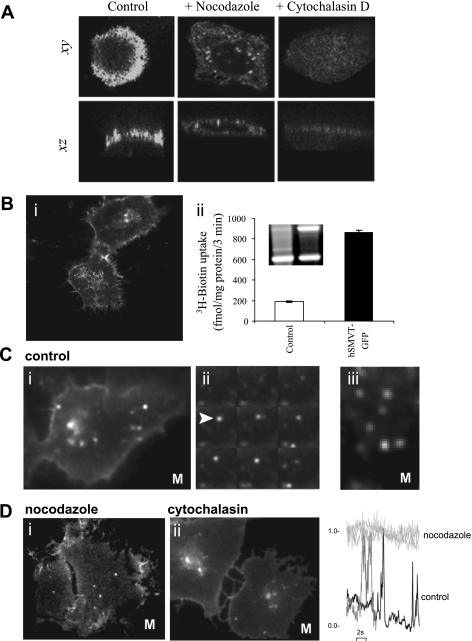
Effect of cytoskeletal disruption on hSMVT trafficking and targeting. A: MDCK cells treated with nocodazole (20 μM, 30 min; middle) or cytochalasin D (10 μM, 30 min; right) before transient transfection were imaged after 18–24 h and compared with transfected control cells (left). Lateral (xy, top) and axial (xz, bottom) confocal planes are shown. B: i, distribution of hSMVT-GFP trafficking vesicles in a stable human-derived duodenal adenocarcinoma cell line (HuTu-80) cell line grown on cover glass-bottomed petri dishes for 5–7 days; ii, [3H]biotin uptake and RT-PCR of hSMVT mRNA (inset) in the stable HuTu-80 cell line. C: i, total internal reflection fluorescence (TIRF) images of hSMVT vesicular dynamics in HuTu-80 cells (at 37°C; Supplemental Movie S1); ii, montage of single frames (taken at 200-ms intervals) of hSMVT vesicles (e.g., arrow) in control cells; iii, lateral motion of hSMVT containing vesicles (Supplemental Movie S2). D: image stills in cells treated with nocodazole (i; Supplemental Movie S3) and cytochalasin (ii; Supplemental Movie S4). Right: examples of fluorescence profile of vesicles in a nocodazole-treated cell (light gray) to illustrate immobility in the TIRF field relative to time 0 and in a control cell (black) and cytochalasin-treated cell (gray) to illustrate transitions into/out of the TIRF field. M, movie.

