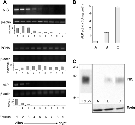
Fig. 2.
NIS mRNA and protein expression along the villus-crypt axis. A: total RNA was extracted from enterocytes sequentially isolated, in nine fractions, from the small intestine along the villus-crypt axis as described in materials and methods. Differential expression of NIS mRNA was analyzed by RT-PCR and standardized with respect to β-actin mRNA expression. Purity of the villus-crypt fraction separation was confirmed by analysis of the expression of alkaline phosphatase (ALP; a villus marker) and PCNA (a crypt marker) mRNAs. Densitometric ratios of NIS, PCNA, and ALP over β-actin expression are shown. B: ALP activity from villus-tip epithelial cells in fractions A (homogenate), B (nonpurified apical membranes), and C (enriched apical membranes). C: NIS immunoblot: lane 1, FRTL-5 cell membranes (10 μg); lanes 2-4, fraction A, B, or C (50 μg). Bottom: ezrin immunoblot as loading control after stripping anti-NIS antibodies. Boxes indicate different gels.

