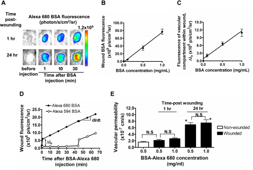
Fig. 1.
Real-time detection of vascular permeability using fluorescent albumin. A: noninvasive fluorescence imaging of BSA-Alexa 680 before and after intravenous (iv) injection at early (1 h) and late (24 h) phase after skin wound. p/s/cm2/sr, photons per square centimeter per steradian. B: topical application of BSA-Alexa 680 directly into the wound. Fluorescence intensity within wound area was measured as a function of BSA titrated into wound. C: in vivo titration of BSA-Alexa 680. BSA-Alexa 680 was injected via tail vein (iv), and fluorescence intensity of BSA within vascular compartment (Io, BSA fluorescence measured 1 min after injection) was measured as a function of injected BSA concentration. D: representative time course of BSA fluorescence within wound. The effect of plasma clearance of circulating BSA-Alexa 680 on permeability measurement was assessed by comparing the response obtained by second dye, in which Alexa 594 BSA was injected 45 min after Alexa 680 BSA injection. E: comparison of baseline vascular permeability in nonwounded intact skin and wounded skin. Vascular permeability is also shown as a function of BSA concentration (0.5 and 1.0 mg/ml) in wounded skin at both 1 h and 24 h postwounding. NS, not significant. *P < 0.05 with respect to 1 h group; n = 3 to 4 mice in each group.

