Fig. 6.
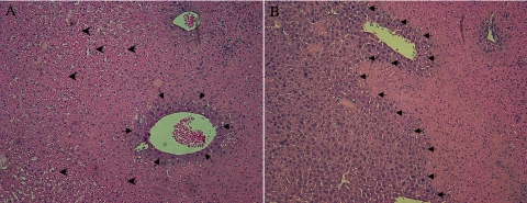
Comparison of the histopathology of liver after partial hepatic IRI in SSAT-wt and SSAT-ko mice. The histology of livers of SSAT-wt (A) and -ko (B) animals subjected to 90 min of ischemia and 12 h of reperfusion were examined (n = 4/group). Histological comparison of uninjured SSAT-wt and -ko liver did not reveal any structural differences (data not shown). Histological assessment of livers from SSAT-wt and SSAT-ko (A and B, respectively) mice after induction of liver injury showed the typical extensive hemorrhagic necrosis and inflammatory infiltrates seen after IRI. However, in livers from SSAT-ko mice (B), there was far less congestion and the extent of necrosis was substantially reduced (small arrows delineate the injured vs. uninjured areas; large arrows indicate the areas of congestion).

