Fig. 5.
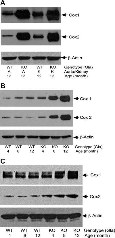
A: COX1 and COX2 in kidney and aortic lysates from 12-mo-old Gla+/0 (WT) and Gla−/0 (KO) mice. Immunoreactivities of COX1 and COX2 in mouse tissues were detected with an anti-COX1 monoclonal antibody (5F6/F4, Abcam, Cambridge, MA) or a monoclonal antibody against COX2 (BD Transduction Laboratories, Lexington, KY). Proteins were reduced in sample loading buffer containing 0.5% β-mercaptoethanol, heated at 80°C for 5 min, separated in SDS-PAGE with a 7–13% gradient, and probed with anti-COX1 and -COX2 antibodies. β-Actin served as internal loading controls. Blots are representative of 3 independent experiments that yielded equivalent results. B: age-dependent accumulation of COX1 and COX2 proteins in aortas of Gla+/0 and Gla−/0 mice. Blots are representative of 3 independent experiments that yielded equivalent results. C: age-dependent accumulation of COX1 and COX2 proteins in mouse aortic endothelial cells isolated from Gla−/0 mice. Proteins in mouse aortic endothelial cell lysates were nonreduced, but they were heated at 80°C for 5 min before they were loaded into SDS-PAGE. β-Actin served as internal loading controls. Results are representative of 3 independent experiments.

