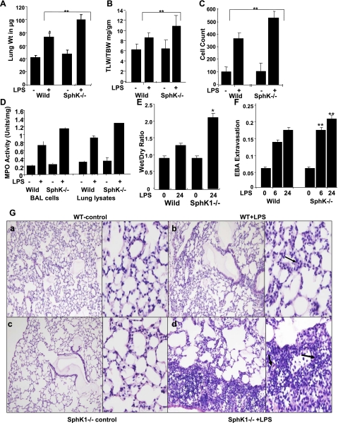
Fig. 4.
Effects of SphK1 deficiency on pulmonary vascular permeability in LPS-treated lungs. Comparison of LPS-induced lung injury in SphK1−/− and WT control mice. A: total wet lung weight was measured. *Significant increase in lung weight in WT and knockout mice. B: total lung weight to body weight (TLW/TBW) compared between WT and knockout mice. *Significant increase. C: right lungs were lavaged using normal saline, and total number of cells in the lavage fluid was measured. D: MPO activity was measured from lung lysates and cells from lavage samples. Average of 3 independent lung lysates was plotted. E: the wet-to-dry lung weight ratio was compared in the WT and SphK−/− mice after LPS-treatment. F: pulmonary vascular leakage was assessed by EBA extravasation assay at 6 and 24 h after LPS was delivered. **Comparison between WT and knockout mice and significant differences. G, a–d: histological comparison of LPS-induced lung injury in WT and SphK1−/− mice. In SphK1−/− mice, LPS produced prominent neutrophil accumulation compared with WT mice (shown by black arrows).

