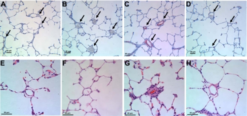
Fig. 3.
In vivo knockdown of HIMF reduces hypoxia-induced histological changes. A–D: lung sections were double-stained with antibodies to α-smooth muscle actin (red) and von Willebrand factor (black). Cell nuclei were counterstained with hematoxylin (blue). Arrows indicate small pulmonary vessels. A: normoxia+Ad-Neg-shRNA. B: normoxia+Ad-HIMF-shRNA. C: hypoxia+Ad-Neg-shRNA. D: hypoxia+Ad-HIMF-shRNA. E–H: hematoxylin and eosin-stained lung sections from rats exposed to normoxia+Ad-Neg-shRNA (E), normoxia+Ad-HIMF-shRNA (F), hypoxia+Ad-Neg-shRNA (G), or hypoxia+Ad-HIMF-shRNA (H) for 14 days. Scale bars = 50 μm.

