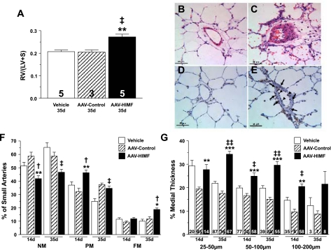
Fig. 6.
Pulmonary gene transfer of HIMF induces vascular remodeling. A: vertical bars represent means ± SE of right ventricular weight/(left ventricular + septal weight) [RV/(LV+S)] in rats 35 days after intratracheal instillation with vehicle, AAV-null (5 × 10¹⁰ VP), or AAV-HIMF (5 × 10¹⁰ VP). The number of animals studied is indicated within each bar. B and C: hematoxylin and eosin-stained lung sections from rats 35 days after being treated with vehicle (B) or AAV-HIMF (5 × 10¹⁰ VP) (C). D and E: lung sections from rats 14 days after being treated with vehicle (D) or AAV-HIMF (5 × 10¹⁰ VP) (E) were probed with antibodies against PCNA (brown) and then counterstained with hematoxylin (blue). F: muscularization of small pulmonary arteries is indicated as non-muscular (NM), partially muscular (PM), or fully muscular (FM) in rat lungs 14 or 35 days after initial intratracheal instillation of vehicle, AAV-null (5 × 10¹⁰ VP), or AAV-HIMF (5 × 10¹⁰ VP). G: comparison of percent medial wall thickness of pulmonary arteries 25–50 μm, 50–100 μm, and 100–200 μm among rats 14 or 35 days after intratracheal instillation of vehicle, AAV-null (5 × 10¹⁰ VP), or AAV-HIMF (5 × 10¹⁰ VP). The number of vessels counted is indicated within each bar. †P < 0.05, ‡P < 0.01, ‡‡P < 0.001 compared with simultaneous vehicle controls. *P < 0.05, **P < 0.01, ***P < 0.001 compared with simultaneous AAV-null.

