
Figure 2. Mechanistic model of microtubule based 1-dimensional diffusion.
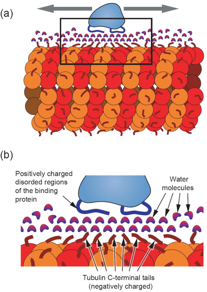
(a) Model of a microtubule binding protein (light blue) diffusing axially along the length of a microtubule (red). The microtubule surface is covered with flexible negatively-charged c-terminal tubulin ends (dark red) [30]. Complementing this, the microtubule binding protein is shown with flexible positively-charged regions (blue) that interact with the microtubule. (b) An expanded view of the boxed area from part (a) depicting a hypothetical interaction scheme in which a layer (or possibly multiple layers) of structured water molecules form an intermediary for the electrostatic interaction. This concept is drawn from the analogous situation of DNA binding proteins in which structural data indicates the existence of water “lubricating” the interfacial surface [40] [37] [38]. This scenario could provide low energy barriers for relatively free sliding along the length of the polymer. An alternative model would be one in which the charged disordered regions of both the microtubule and binding protein interact directly with very little interfacial water. In either scenario, the non-specificity of the interaction is a key feature for providing low energy barriers and the potential for fast sliding.