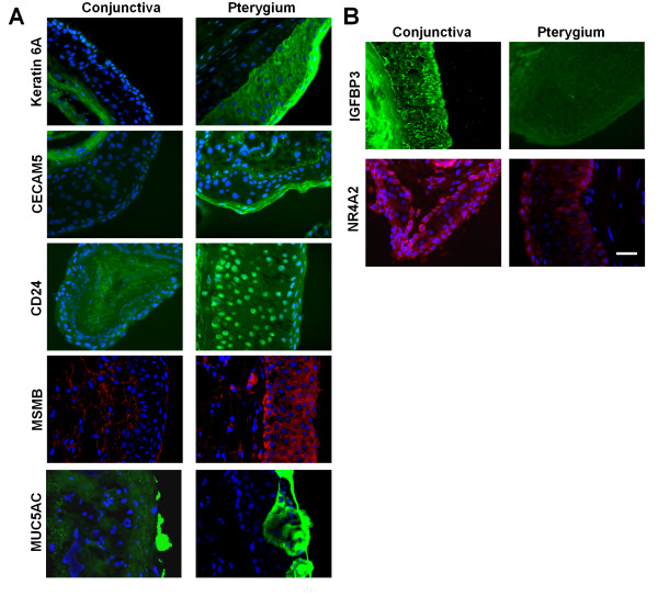
Figure 8.
Tissue localisation of important molecules in pterygium. Immunohistochemical staining images of uninvolved conjunctiva and pterygium tissue. Nuclear position (and indirectly, tissue architecture) was shown using counter-staining with DAPI, in blue. The same magnification was used in all images: Scale Bar = 100 micrometers. A. Proteins with increased expression in pterygium. Staining for keratin 6A, CECAM5, CD24 and MUC5AC was shown in green and MSMB, in red. B. Proteins with decreased expression in pterygium. Staining for IGFBP3 and NR4A2 was shown in green and red respectively.

