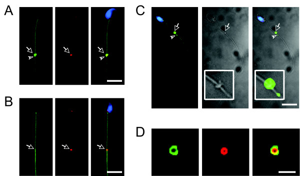
Figure 1.
Transient localization of DNAJB13 to sperm annulus. In addition to the axonemal localization, DNAJB13 (green) was also co-localized with an annulus constituent SEPT4 (red) to the sperm annulus (arrows) in developing spermatids (A), but not in mature spermatozoa (B). The phase contrast images of a spermatid further showed that the localization of DNAJB13 to the annulus (see insets in the phase contrast images) (C). Note the DNAJB13 antibody also labeled a small dot (arrowheads) below the annulus (A, C). The sperm nuclei were stained with DAPI (blue). The scale bar represents 10 μm; (D) Images of an annulus released from a damaged spermatid. The scale bar represents 2 μm.

