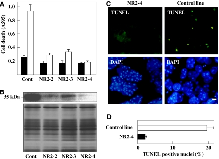
Figure 3.
Suppression of HR cell death in OsNAC4 RNAi transformants. (A) Cell death detected by Evans blue staining in OsNAC4 RNAi transformant lines (NR2-2, NR2-3 and NR2-4) before (solid column) and 8 h after (open column) inoculation with the avirulent N1141 strain. (B) Accumulation of OsNAC4 was detected with an anti-OsNAC4 antibody in OsNAC4 RNAi knock-down lines. Proteins (10 μg) from vector transformed cells (Cont) and RNAi transformant lines were separated on a 12.5% (w/v) SDS–PAGE and transferred to a nitrocellulose membrane. Then OsNAC4 were detected by immunoblotting with an anti-OsNAC4 antibody (top part). The same amount of each fraction was separated by SDS–PAGE and proteins were detected by silver staining (lower part). (C) DNA fragmentation in OsNAC4 knock-down (left panels) and control (right panels) lines was assessed after inoculation with the avirulent N1141 strain. TUNEL (upper panels) and DAPI (lower panels) staining are shown. Bar represents 10 μm. (D) Percentage of TUNEL-positive nuclei in NR2-4 and vector transformed lines (control line) inoculated with N1141. The percentage of TUNEL-positive nuclei was determined by counting nuclei within 20 individual fields. Each determination was done with at least 2000 nuclei in each of three independent experiments. A full colour version of this figure is available at The EMBO Journal online.

