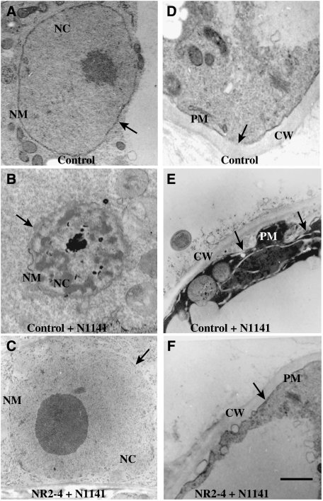
Figure 4.
Morphological analysis using transmission electron microscopy. (A) A nucleus in a cell transformed with empty vector (control cell) 12 h after water treatment. The arrow indicates a normal nucleus. (B) A nucleus in empty vector-transformed cells (control cell) 12 h after inoculation with the avirulent N1141 strain. The arrow indicates condensed nucleus. (C) A nucleus in the OsNAC4 knock-down NR2-4 line after a 12-h inoculation with the avirulent N1141 strain. The arrow indicates a normal nucleus. (D) The cell wall and plasma membrane in a cell transformed with empty vector (control cell) 12 h after water treatment. The arrow indicates a normal plasma membrane. (E) The cell wall and plasma membrane in empty vector transformed cells (control cell) 12 h after inoculation with the avirulent N1141 strain. The arrows indicate the separated-plasma membrane from the cell wall. (F) The cell wall and plasma membrane in the OsNAC4 knock-down NR2-4 line after a 12-h inoculation with the avirulent N1141 strain. The arrow indicates a normal plasma membrane. CW, cell wall; NC, nucleus; NM, nuclear membrane; PM, plasma membrane. Bar represents 1 μm.

