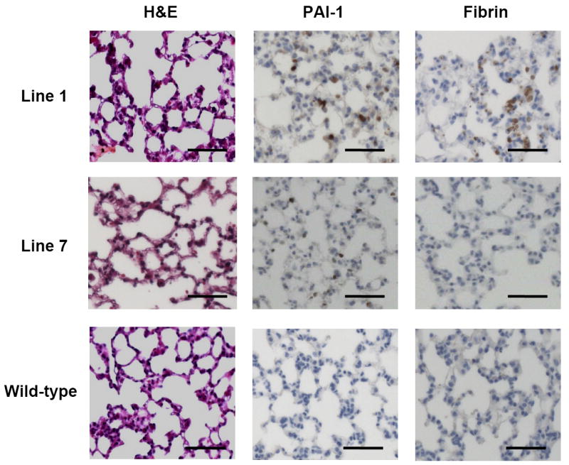
Figure 5. Histology of transgenic pulmonary tissue.

Hematoxylin and eosin staining, fibrin immunohistochemistry, and PAI-1 immunohistochemistry are shown for pulmonary tissue from one N1 transgenic mouse from each of lines 1 and 7, and a wild-type mouse from line 1, at 20X magnification. Bars indicate 50 μm.
