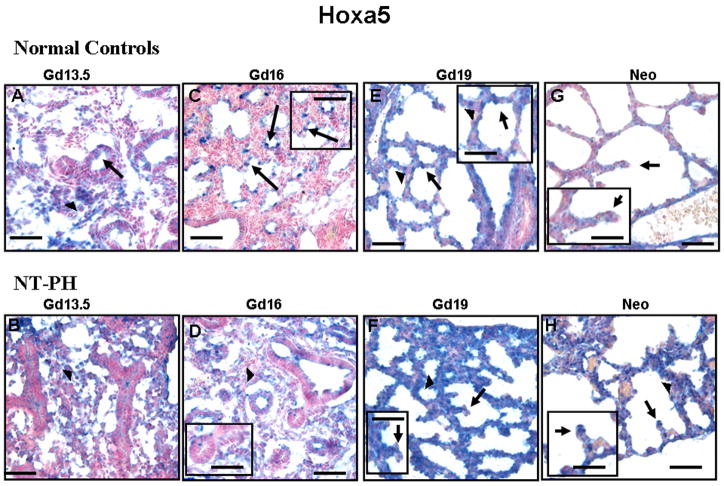
Figure 3. Hoxa5 protein immunolocalization in normal developing mouse lung and in NT-PH lungs. Bar = 50μ in Panels A–H and 20μ in corresponding inserts.
In Gd13.5 normal controls (A) and NT-PH (B) lungs, Hoxa5 protein was mostly localized to mesenchyme (arrowheads) with minimal expression in epithelial cells of columnar-lined airways (arrows). In Gd16 normal lungs (C), Hoxa5 protein localization changed being mostly restricted to epithelial cells of branching airway tips (arrows) (see higher magnification insert). This change was not seen in Gd16 NT-PH lungs where Hoxa5 remained mostly diffusely expressed in mesenchyme (arrowheads). Hoxa5 protein remained mostly localized to epithelial cells (arrows) in Gd 19 normal controls (E). Gd19 NT-PH lungs (F) also had persistent expression in thicker mesenchyme (arrowheads) whereas epithelial cells (arrows) especially at septal tips remained negative. In neonatal normal controls (G), Hoxa5 epithelial cell expression was less intense as compared to strong Hoxa5 mesenchymal (arrowheads) and epithelial expression (arrows) especially at septal tips in hypoplastic lungs (H).

