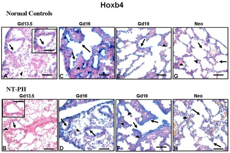
Figure 4. Hoxb4 protein immunolocalization in normal developing mouse lung and in NT PH lungs, Bar = 50μ in Panels A–H and 20μ in corresponding inserts.
Hoxb4 protein was localized to both mesenchymal (arrowheads) and epithelial cells (arrows) in Gd13.5 normal controls (A) and NT-PH lungs (B) but epithelial cell expression was more intense. Gd16 normal controls (C) and NT-PH lungs (D) had strong epithelial cell expression, but Hoxb4 mesenchymal cell (arrowheads) expression was more intense in NT-PH lungs than in the normal controls. Gd19 normal controls (E) and NT-PH lungs (F) had similar Hoxb4 mesenchymal (arrowheads) and epithelial (arrows) cell localization, but NT-PH lungs had increased intensity of Hoxb4 in epithelial cells (arrows), especially in bronchiolar airways as well as somewhat increased mesenchymal expression (arrowheads). Compared to normal neonatal lungs (G), epithelial cell (arrow) Hoxb4 staining remained stronger in neonatal NT-PH lungs (H). Thicker mesenchyme (arrowhead) in NT-PH lungs continued to have greater intensity of Hoxb4 positive cells than seen in the normal lungs.

