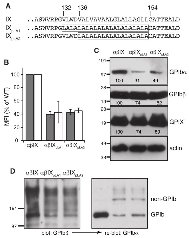
Fig. 3. Mutational effect on complex expression and assembly by replacement of the GPIX TM domain with pLA sequences.
(A) TM sequences in wild type and domain replacement GPIX constructs. The pLA sequence in each mutant is boxed. (B) Surface expression levels of GPIbα (gray) and GPIX (white) in domain replacement cells. Measurement and quantitation followed the description in Fig 1B. (C) Overall expression of GPIbα and GPIX. Cell lysates were resolved in reducing SDS gels. The relative band intensity is the average of 3 independent experiments. (D) GPIb formation in domain replacement cells. Cell lysates were resolved in non-reducing SDS gels and probed as indicated. The blots are representatives of 2 independent experiments.

