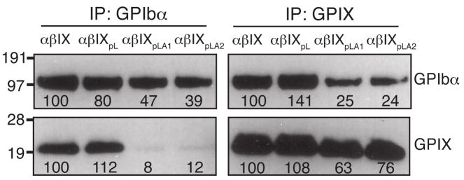
Fig. 4. Replacement of the GPIX TM domain with pLA sequences, but not the pL sequence, weakened the interaction between GPIb and GPIX.
Cell lysates were co-immunoprecipitated with antibodies AK2 or FMC25, resolved in a reducing SDS gel, and immunoblotted with WM23 or anti-IX polyclonal antibody. Intensities of the GPIbα and GPIX bands were quantitated by densitometry and expressed as a percentage of the wild type level. The numbers below the bands are the average of 3 independent experiments.

