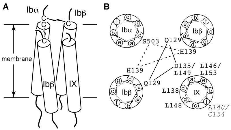
Fig. 9. A four-helical bundle model for the TM domains of the GPIb-IX complex.
(A) The side view of the TM bundle. Each TM helix is shown as a rod traversing the membrane bilayer. (B) The helical wheel diagram of the TM bundle. The possible interactions among the polar residues are marked by lines. The residues in GPIX that play an role in modulating GPIb formation, as well as A140 and Cys154 (Italic), are marked.

