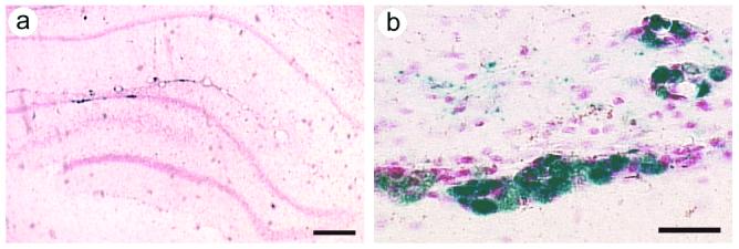
Figure 5.

Recombinant gene expression in the dentate gyrus. After the water maze training was completed, 3-4 weeks after gene transfer, the rats were sacrificed, and ß-gal was detected with X-gal staining. These vectors coexpress PkcΔ (or PkcΔGG) and ß-gal. (A) A low power view from a rat that had received PkcΔ, shows X-gal positive cells in the dentate granule cell layer. The section was counterstained with neutral red. (B) A high power view showis X-gal positive cells with neuronal morphology in the dentate granule cell layer; some X-gal positive cells are also present outside of the granule cell layer. Scale bars: (A) 300 μm; (B) 30 μm.
