
Fig. 5.
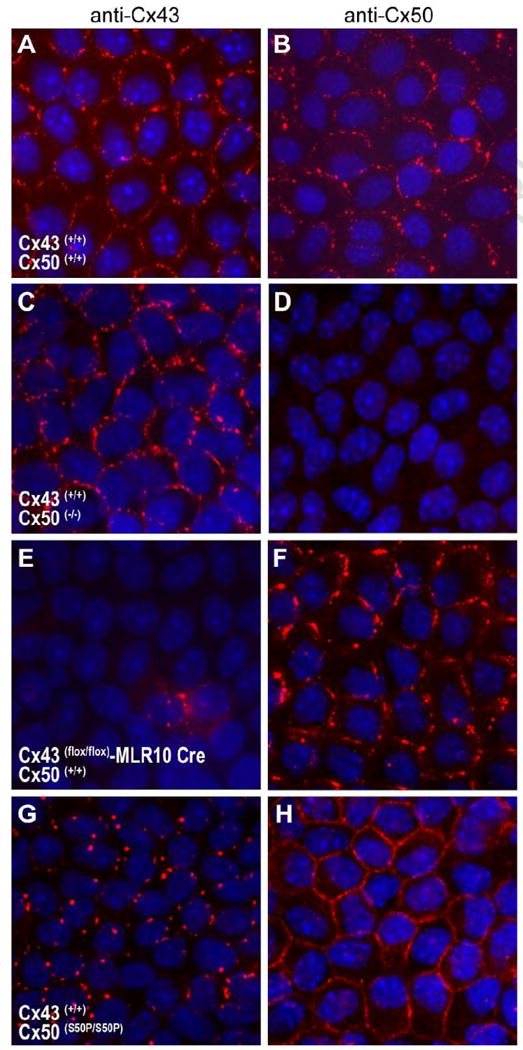
Immunofluorescent staining of the Cx43 and Cx50 lens proteins in lens epithelial cells. Lens capsules from wild-type (A, B), Cx50 knockout (C, D), conditional Cx43 knockout (E,F) and homozygous Cx50-S50P mutant (G,H) lenses were immunostained with antibodies raised against Cx43 and Cx50 and examined by fluorescence microscopy. Merged images show Cy3 staining of connexins (red) and DAPI staining of cell nuclei (blue). Discernable aberrations in endogenous connexin 43 staining were visible in the presence of mutant Cx50-S50P (G) a phenomenon not seen in the Cx50 knockout animal (C).