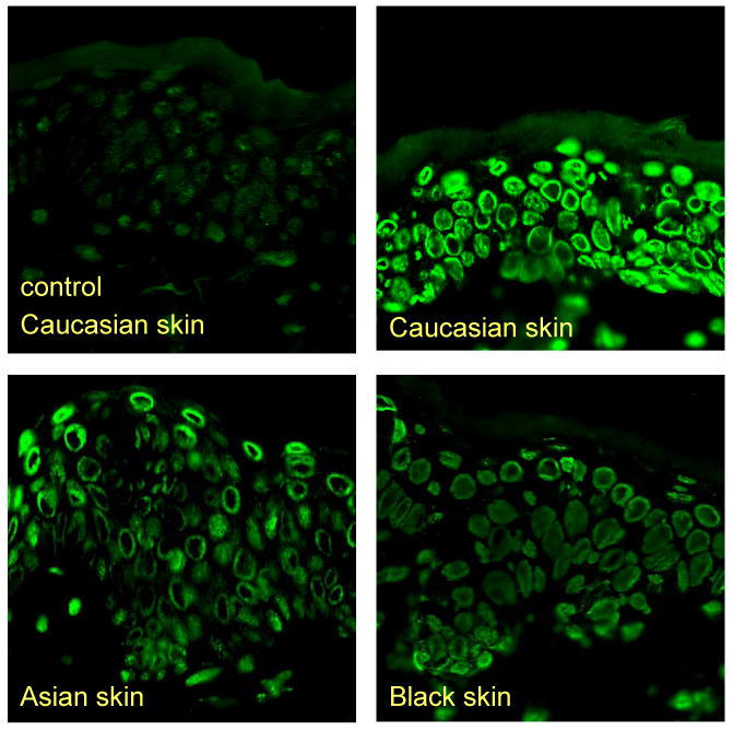
Figure 2. CPD in Skin of Different Ethnicity.

Immunohistochemical detection of CPDs in skin of different ethnicities immediately after UVR. The fluorescence detection technique uses the binding of FITC-labeled antibodies that are reactive with DNA photoproducts. A considerably higher amount of CPDs is detected in White skin compared to Asian or Black skin.
