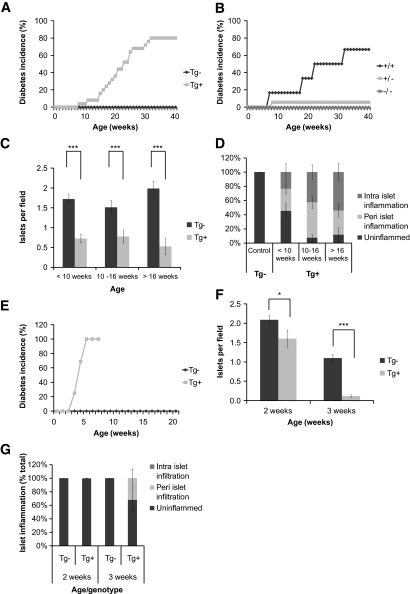
FIG. 7.
IL-21Tg mice develop spontaneous type 1 diabetes on the C57Bl/6 background, the severity of which is increased in the context of NOD alleles. A: IL-21Tg mice and wild-type littermate controls. Diabetic incidence was calculated per group as two consecutive readings over 250 mg/dl and displayed as percentage of diabetic mice per group (Tg−, n = 24; Tg+, n = 25). Diabetes incidence was scored as survival curve data and is different by log-rank test (P < 0.0001). B: Weekly measurements of blood glucose were performed on IL-21Tg mice after crossing to the IL-21R knockout (C57BL/6), and diabetes incidence was calculated (all mice Tg+, IL-21R+/+, n = 7; IL-21R+/−, n = 25; IL-21R−/−, n = 8). Paraffin-embedded pancreatic tissue from wild-type and IL-21Tg mice was sectioned, stained with an anti-insulin polyclonal antibody, and counterstained with hematoxylin. C: Total number of insulin-positive islets per visual field. D: Individual islets were scored for the presence of peri- and intra-islet infiltration and displayed as percentage of infiltrated islets per group (<10 weeks, Tg−, n = 3; Tg+, n = 8; 10–16 weeks, Tg−, n = 12; Tg+, n = 14; >16 weeks, Tg−, n = 7; Tg+, n = 12). Original magnification: ×10. E: IL-21Tg mice on the C57Bl/6 background were crossed with the NOD strain to generate a B6×NOD F1 mixed background. Weekly measurements of blood glucose were performed on IL-21Tg littermate controls, and diabetes incidence was calculated (Tg−, n = 19; Tg+, n = 16). Diabetes incidence was scored as survival curve data and is different by logrank test (P < 0.0001). Paraffin-embedded pancreatic tissue from this cross was quantitated for total number of insulin-positive islets per visual field (F) and individual islets scored for the presence of peri- and intra-islet infiltration and displayed as percentage of infiltrated islets per group (G) (2 weeks, Tg−, n = 11; Tg+, n = 12; 3 weeks, Tg−, n = 15; Tg+, n = 7).

