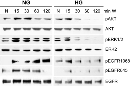
FIG. 5.
Weakening of wound induced EGFR signaling pathways in human corneal epithelial cells cultured in high glucose. THCE cells were cultured in normal glucose (NG; 5 mmol/l d-glucose) and high glucose (HG; 25 mmol/l d-glucose) for 48 h and starved overnight in keratinocyte basal medium with the same glucose conditions as before. Cells were wounded extensively by sequence comb scratching (W) at the indicated times (N, without wound). Cells were then lysed, and equal amounts of proteins of 10–30 μg were subjected to Western blot using antibodies against phosphorylated proteins pAkt, pERK1/2, or pEGFR (Y845, -Y1,068). After stripping off the immunoreactivities, the membranes were reprobed with antibody against Akt, ERK2, or EGFR, respectively, for proper protein loading. As each condition was processed separately for Western blotting, each comparison between the control and treated samples were made only within each group.

