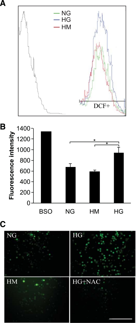
FIG. 6.
Overproduction of ROS in cultured primary human corneal epithelial cells. Primary human corneal epithelial cells were cultured in medium containing normal glucose (NG), high glucose (HG), or high mannitol (HM) or medium with BSO (250 μmol/l) as positive control for 24 h and then incubated with cell-permeable DCFH-DA (5 μmol/l) for 45 min. A: Cells were detached by trypsin digestion and washed with PBS. Fluorescence of oxidized DCFH-DA by intracellular ROS was detected by flow cytometry for quantitative measurement. Fluorescence peaks in normal glucose, high mannitol, or high glucose were compared in terms of DCF-positive (DCF+) cells relative to the cells with nonfluorescence peak on the left. B: The mean fluorescence intensity of flow cytometry was quantified as the means ± SD. *P < 0.05. C: THCE cells were grown in normal glucose, high mannitol, or high glucose with or without NAC for 24 h and incubated with DCFH-DA as mentioned above. ROS generation was visualized under a fluorescent microscope. Scale bar = 50 μm. (A high-quality digital representation of this figure is available in the online issue.)

