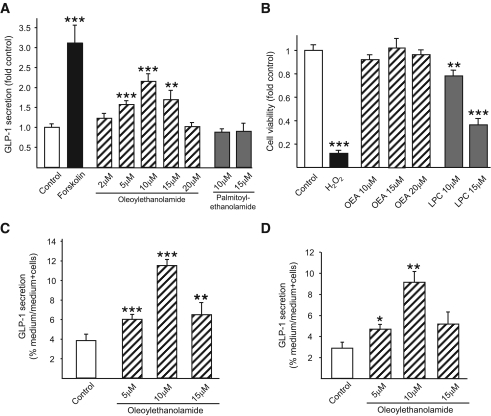
FIG. 2.
Effects of OEA on GLP-1 secretion. mGLUTag cells (n = 9–12) (A), hNCI-H716 cells (n = 8) (C), and FRIC cells (n = 4) (D) were incubated with medium alone (1% DMSO, negative control), forskolin (10 μmol/l, positive control), OEA (2–20 μmol/l), or PEA (10–15 μmol/l, negative control) for 2 h. GLP-1 content of media and cells was determined by RIA. B: To determine potential effects on cell viability, mGLUTag cells were incubated with medium alone (1% DMSO, negative control), H2O2 (5 mmol/l, positive control), OEA (10–20 μmol/l), or LPC (10–15 μmol/l) for 2 h, followed by MTT assay (n = 8–16). *P < 0.05; **P < 0.01; ***P < 0.001 vs. control.

