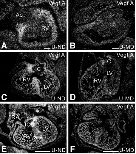
FIG. 5.
Diabetes-induced decrease in cardiac VEGF expression as shown by in situ hybridization. In the E14-ND offspring, VEGF is abundantly expressed in the myocardium aligning the cushions of the outflow tract of the heart (A), in the ventricular septum (C), and in the myocardium around the cushions of the inflow tract of the heart (C and E). At E14, we see a diabetes-induced decrease in VEGF expression in all L-MD embryos (n = 2) and in 75% of the four-unit MD embryos (n = 4) in the myocardium aligning the cushions of the outflow (B) and inflow tract of the heart (D and F), in the ventricular septum (D). Ao, aorta; IC, inflow tract cushion; LV, left ventricle; RA, right atrium; RV, right ventricle; SV, sinus venosus. Scale bar = 200 μm.

