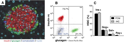
FIG. 1.
Pancreatic islet hormone-secreting cell subsets as assessed by immunofluorescence and flow cytometry. A: Pancreas section from a naïve C57BL/6 mouse simultaneously stained for the three major endocrine cell subsets: insulin positive (β-cells, red), glucagon positive (α-cells, green), and somatostatin positive (δ-cells, dark blue). Nuclei are shown in turquoise. Magnification ×400. B: Islet endocrine cells by flow cytometry. Handpicked pancreatic islets isolated from one naïve C57BL/6 mouse were dissociated into a single-cell suspension and intracytoplasmically stained for insulin (red), glucagon (green), and somatostatin (blue). All other islet components (e.g., other endocrine, endothelial cells) and residual exocrine cells are depicted in gray. For presentation purposes, insulin-, glucagon-, and somatostatin-positive endocrine cell subsets (typically making up ∼80% of healthy islets) were normalized to 100%. C: Quantitative analysis of islet cell subsets of naïve C57BL/6 mice by flow cytometry (■) and immunofluorescence (▩). For flow cytometry, >10,000 events of dissociated and stained islet cells per mouse were acquired. Frequencies (means ± SE, n = 5 mice) were: insulin positive 73.5 ± 2.4%, glucagon positive 19.2 ± 1.9%, somatostatin positive 5.5 ± 0.6%, and pancreatic polypeptide positive 1.7 ± 0.5%. For immunofluorescence, the three major endocrine cell subsets were quantified by independent, triplicate, or quadruplicate scorings (>350 endocrine cells were counted of at least 12 randomly selected islets). Their frequencies (means ± SE, n = 3 mice) were 76.7 ± 1.1%, 17.3 ± 3.3%, and 5.9 ± 4.0% for insulin-, glucagon-, and somatostatin-positive cells, respectively. Gcg, glucagon; Ins, insulin; Ppy, pancreatic polypeptide; Sst, somatostatin. (A high-quality digital representation of this figure is available in the online issue.)

