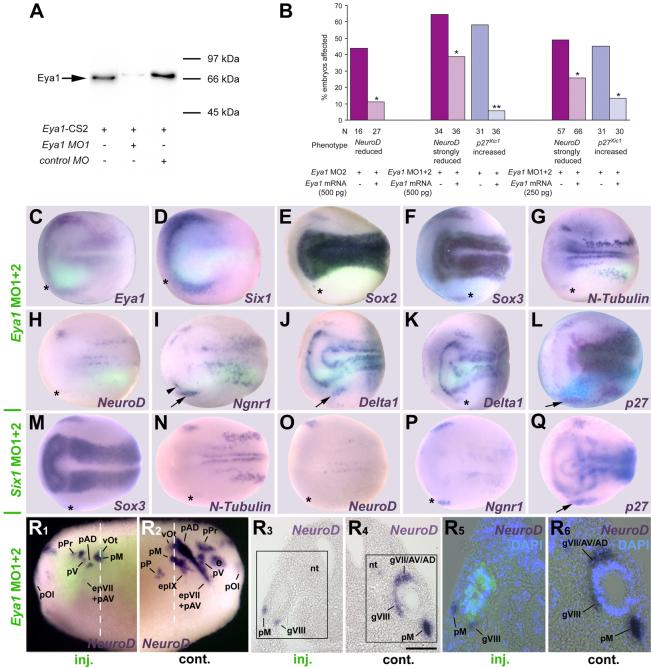
Fig. 2.
Effects of Eya1 and Six1 knockdown on markers of neurogenesis and placodal ectoderm. A: Immunoblot showing that Eya1MO1 but not control MO blocks synthesis of Eya1 protein. B: Co-injecting Eya1MO2 or Eya1MO1+2 with myc-tagged Eya1 mRNA significantly restores NeuroD and prevents ectopic p27Xic1 expression (X2 test; *: p< 0.05, **: p< 0.001) in three independent rescue experiments. C-Q: Neural plate stage embryos after unilateral injection (lower half) of Eya1MO1+2 (C-L) or Six1MO1+2 (M-Q). In all cases, where myc-GFP was coinjected as lineage tracer (C-K), embryos are shown superimposed with green fluorescent channel. Asterisks indicate reductions, arrows and arrowheads indicate increased marker gene expression. For Delta1 after Eya1MO1+2 injections two different phenotypes are depicted (J, K). R: Tail bud stage embryo after unilateral injection (R1, R3, R5: injected side; R2, R4, R6: control side) of Eya1MO1+2 reveals reduction of NeuroD expression in all neurogenic placodes or derivative ganglia. R3 and R4 depict a section at the level indicated (white line) with boxed areas shown magnified in R5 and R6, respectively, superimposed with green (myc-GFP co-injected with Eya1MO1+2) and blue (DAPI) fluorescence. Residual NeuroD expression is confined to cells receiving little or no MO. Bar in R4: 100 μm (for R3, R4). Abbreviations: e: eye; epVII+AV: facial epibranchial and anteroventral lateral line placode; epIX: glossopharyngeal epibranchial placode; gVII/AV/AD: ganglion of the facial, anteroventral and anterodorsal lateral line nerve; gVIII: vestibulocochlear ganglion; nt: neural tube; pAD: anterodorsal lateral line placode; pM: middle lateral line placode; pOl: olfactory placode; pP: posterior lateral line placode; pPr: profundal placode; pV: trigeminal placode; vOt: otic vesicle.

