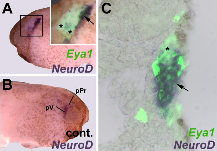
Fig. 8.
Cell-autonomous effects of Eya1 on placodal neurogenesis. Panels show distribution of NeuroD in grafts of placodal ectoderm from embryos injected with Eya1 and myc-GFP mRNA to uninjected hosts. Boxed area is magnified in insert of A. Insert of A and C are shown superimposed with green fluorescent channel to reveal myc-immunostaining. While NeuroD expression in profundal (pPr) and trigeminal (pV) placodes is reduced in the graft (asterisk, A, C) compared to control side (B), NeuroD is expressed ectopically (arrow) at graft border but not in host ectoderm (A, C).

