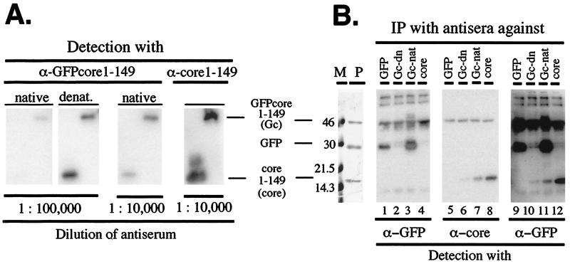
Figure 4.
Efficient antibody induction by GFPcore1–149. (A) Reactivity of anti-GFPcore1–149 antisera in Western blots. Poly(vinylidene difluoride) membranes containing GFPcore1–149 and core1–149 protein (5 ng per lane) were separately incubated with the rabbit antisera elicited against native particulate or denatured GFPcore1–149 protein at the indicated dilutions. A polyclonal serum against core1–149 protein served as control. Antigen-bound antibodies were visualized by using anti-rabbit-IgG-peroxidase conjugates and a chemiluminescent substrate. (B) Immunoprecipitation of native antigens by anti-GFPcore1–149 antisera. Native GFPcore1–149 (Gc) and core1–149 (core) particles and eGFP (GFP) were incubated with immobilized antibodies against native or denatured GFPcore1–149, against native GFP, and against core1–149 protein. Precipitated proteins were detected by Western blotting using monoclonal anti-GFP (lanes 1–4) or polyclonal anti-core antibodies (lanes 5–8) as in A. Lanes 9–12 show the anti-core blot after reprobing with anti-GFP antibodies. The left panel shows a Coomassie blue-stained SDS/PAGE gel of the input solution (lane P) alongside protein size markers (lane M). Note the efficient precipitation of native GFP by the serum against native (lanes 3 and 11) but not denatured (lanes 2 and 10) GFPcore1–149.

