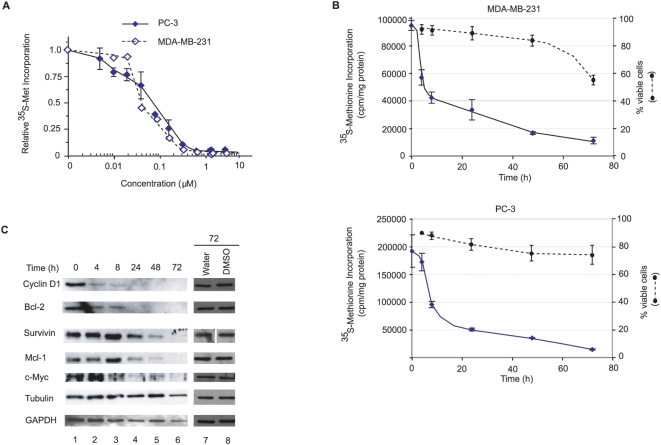
Figure 3. Silvestrol inhibits translation in vivo in MDA-MB-231 breast and PC-3 prostate cancer cells.
A. Relative rate of 35S-Met incorporation in MDA-MB-231 breast and PC-3 prostate cancer cells as a function of silvestrol concentration. Cells were exposed to the indicated concentrations of silvestrol for 1 h in Met-free DMEM with 10% dialyzed FBS, during which the last 15 min, 35S-Met was added. Results are the average of duplicates with the error of the mean shown. Values are standardized against total protein content and plotted relative to DMSO controls, which were ∼100,000 and 400,000 cpm/µg for MDA-MB-231 and PC-3 cells, respectively. B. Kinetics of protein inhibition and cell death following exposure to silvestrol. Cells (60,000 per well in a 24-well plate) were exposed to 25 nM silvestrol for the indicated periods of time. One set of cells was used to quantitate protein synthesis following 35S-Met-labeling, TCA precipitation, and scintillation counting. A parallel set of dishes (200,000 cells per well in a 6-well plate) was used to measure the percentage of viable cells by Annexin V/P.I. staining followed by FACs analysis. These values were normalized to those obtained in the presence of vehicle (1% DMSO), which was set to 100%. C. Reduction in expression of eIF4E-responsive gene products in MDA-MB-231 cells exposed to silvestrol. Western blotting was used to evaluate protein levels from cell lysates harvested at the indicated time points. The panels for survivin (lanes 7 and 8) were analyzed on the same gel, but not on adjacent lanes.

