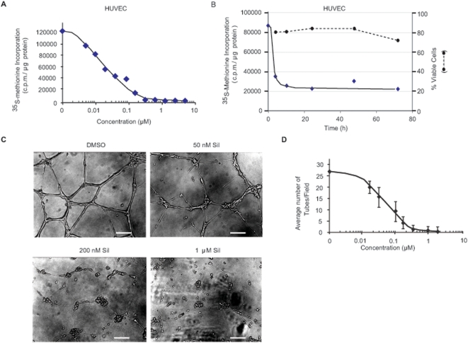
Figure 5. Silvestrol inhibits protein synthesis and suppresses endothelial cell tube formation.
A. The relative rate of 35S-Met incorporation in HUVECs as a function of silvestrol concentration. Cells were exposed to the indicated concentrations of silvestrol for 1 h, and in the last 15 min, 35S-Met was added to the cells. Extracts were prepared and the amount of TCA-insoluble 35S-Met determined. Results are the average of duplicates with the error of the mean too small to be seen. Values are standardized against total protein content. B. Kinetics of protein inhibition versus cell death following exposure of HUVECs to silvestrol. HUVECs were exposed to 25 nM silvestrol for the indicated periods of time. One hour before the end of treatment, media was removed, cells washed with PBS and incubated for an additional hour with silvestrol in Met-free DMEM. For the last 15 min, cells were labeled with 35S-Met, followed by TCA precipitation and scintillation counting. A parallel set of dishes (200,000 cells/well in a 6-well plate) were used to measure the percentage of viable cells by Annexin V/P.I. staining and FACs analysis. These values were normalized to those obtained in the presence of 1% DMSO, which was set at 100%. These values are plotted on the right ordinate and as a dashed line. C. Disruption of tube formation by HUVECs in the presence of silvestrol. HUVECs were seeded on BD Matrigel™ Matrix basement membrane (BD Biosciences, Bedford MA) in 24-well plates in triplicate in the presence of increasing concentrations of silvestrol and 24 h later monitored for tube formation. Photographs were taken with a Nikon eclipse TE300 microscope. The bar at the bottom of each photograph corresponds to 50 µm D. Quantitation of tubules (chord-like structures) observed per field. A total of 15 different fields were used for each data point and the errors represent the standard deviation.

