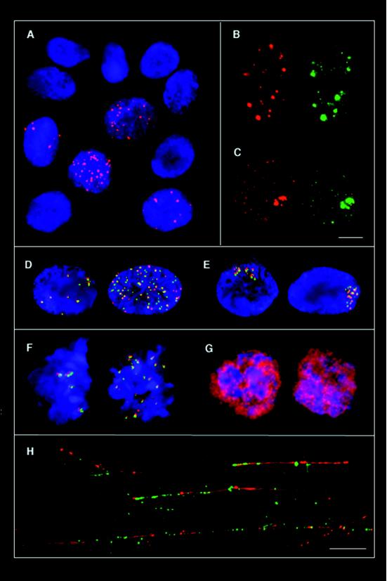
Figure 1.
Association of ssDNA foci with recombination proteins Rad51 and RPA. (A) Visualization of ssDNA foci in human PPL fibroblasts 1 hr after 60Co irradiation with a dose of 10 Gy. Nuclear foci containing ssDNA are stained by red anti-BrdUrd antibody. Nuclei are counterstained with 4′,6-diamidino-2-phenylindole. At this time point, no Rad51 foci are seen. (B and C) Colocalization of Rad51 protein and ssDNA foci in PPL cells at 6 hr (B) and 24 hr (C) after irradiation. Nuclei on the left show ssDNA staining (red) and the nuclei at the right anti-Rad51 immunofluorescence (green) of the same cells. (D and E) Colocalization of replication protein A detected by anti-RPA antiserum (green) and ssDNA foci (red) at 6 hr (D) and 24 hr (E) after irradiation. To facilitate the demonstration of colocalizations, the green signals were shifted purposely by one pixel to the right and by one pixel to the bottom. (F) Colocalization of Rad51/DMC1 (green) and ssDNA (red) in mouse meiotic prophase cells. (G) As a control to demonstrate the uniform labeling of meiotic DNA with BrdUrd, anti-BrdUrd staining was performed on denatured spermatogenic cells. (H) Colocalization of Rad51 (green) and ssDNA (red) on experimentally stretched chromatin fibers from a γ-irradiated culture. Because individual cells are completely destroyed during preparation, the ssDNA fibers from the optical field shown do not necessarily represent the DNA from one cell. (Bars in C and H correspond to 10 μm.)

