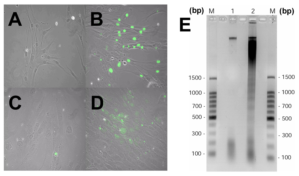
Figure 1.
Fluorescence Images of treated cells were observed on a Zeiss Axioskop microscope and pictures were taken using a charged coupled device (CCD) camera, MagnaFire, Model S99806 (Olympus American) at a magnification of X200. (A) Image of HBVECs treated with 100µl/ml (v/v) RBC-SUP. (B) HBVECs (nucleus stained green) treated with 100µl/ml (v/v) pRBC-SUP undergoing apoptosis. (C) Neuroglia cells treated with 100µl/ml (v/v) RBC-SUP. (D) Neuroglia treated with 100µl/ml (v/v) pRBC-SUP undergoing apoptosis. (E) DNAs from HBVECs extracts were resolved by agarose gel electrophoresis, stained with ethidium bromide and visualized by exposure to UV light. Images were taken using FD67110 Fotodyne FOTO/Analyst Luminary Workstations Systems (Fotodyne, Inc., Hartland, WI.) and the negative image was generated via Adobe Photoshop Software 5.0 and formated with Adobe Illustrator 10.0 software. The results are typical representations of at least two independent experiments. The lanes are as follows: DNA ladder marker (lane M), RBC-SUP (lane 1) and pRBC-SUP (lane 2).

