
Figure 5.
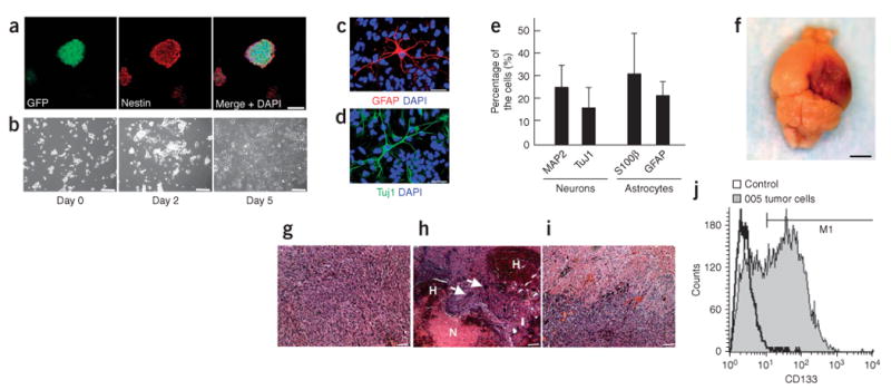
005 tumor cells show the characteristics of BTICs. (a) Confocal microscopic analysis of neurosphere-like structures formed by 005 tumor cells. Cells stained with nestin-specific antibody are shown in the middle panel. A merged image with DAPI staining is shown in the right panel. Scale bar, 50 μm. (b) Phase-contrast images of 005 tumor cells showing elongated processes in response to serum addition in the medium. Images were taken on days 0, 2 and 5 after the serum addition. Scale bars, 50 μm. (c–e) 005 tumor cells expressing neuronal or astrocyte markers. Five days after the addition of FBS to the medium (final 10% FBS), cells were fixed and stained with GFAP-specific antibody (c) or Tuj1-specific antibody (d) and observed by confocal microscopy. Merged mages with DAPI are shown. Scale bars, 30 μm. The proportions of cells positive for neuronal or astrocyte marker are shown in e. Data represent means ± s.d. (f) The gross appearance of tumor formation in the brain of a NOD-SCID mouse after injection of 100 of 005 tumor cells. Scale bar, 3 mm. (g–i) Histological characteristics of the tumor caused by the injection of 005 tumor cells in NOD-SCID mice, including increased cellular density (g), intratumoral hemorrhage (indicated by H in h), necrosis (indicated by N in h), giant cell formation (indicated by white arrows in h) and infiltrative characteristics (i). Scale bars, 50 μm. (j) FACS analysis of 005 tumor cells in culture incubated with CD133-specific antibody. Unstained cells were used as a control.