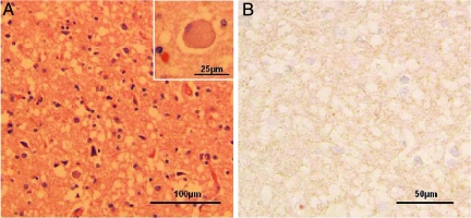
Figure 1.
Histology and immunohistochemistry of brain tissues. A: Widespread spongiform degeneration, often with large vacuoles, was observed in the cortex. Reactive astrocytes also were prominent. Inset: Ballooned neuron. B: Immunostaining for the prion protein demonstrated very minute positive granules, some of which are indicated with circles, in the cortical regions with severe spongiform degeneration using antibody 3F4.

