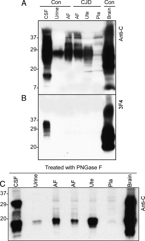
Figure 4.
Comparison of profiles of PrP from different tissues. A: The profile of PrP from the uterus, placenta, and amniotic fluid (AF) was compared with that of PrP from brain, CSF, and urine using Western blot analysis with anti-C antibody. The migration of most PrP species from the uterus, placenta, and AF is between 24 and 34 kDa, significantly different from that of PrP from the brain and CSF but similar to that of urine PrP. B: PrP from CSF, urine, amniotic fluid (AF), uterus (Ute), placenta (Pla), and brain was probed with 3F4. PrP was detectable in CSF and brain samples but not in urine, AF, uterus, and placenta. C: Samples from CSF, urine, AF, uterus, placenta, and brain were deglycosylated with PNGase F before Western blot analysis with anti-C antibody. As expected, PrP from CSF and brain shows two major bands corresponding to the ∼28- to 29-kDa full-length and ∼18- to 19-kDa N-terminally truncated PrP whereas PrP from urine, AF, uterus, and placenta shows only one major band corresponding to the ∼18- to 19-kDa N-terminally truncated PrP fragment.

