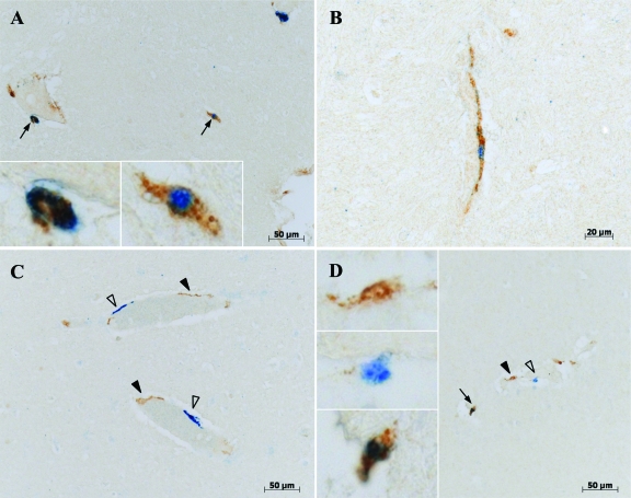
Figure 5.
Phenotype of EGFP+ cells in the brain. Double-label immunohistochemistry of brain tissues (A–B: cortex; C–D: cerebellum) clearly shows three populations of cells. The open arrowheads point EGFP single-positive cells (blue, vector blue chromogen), black arrowheads point CD163 single-positive perivascular macrophages (brown, DAB chromogen), and black arrows point EGFP/CD163 double-positive cells (blue/brown). Insets show higher magnification of single or double positive cells.

