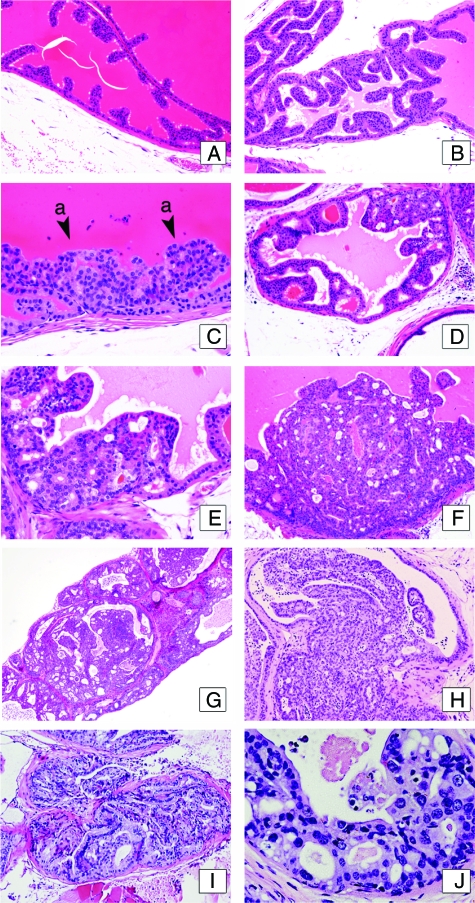
Figure 1.
Histology of lesions of the anterior prostate. Sections of 6- to 12-month-old mice. A: Anterior prostate from wild-type mouse showing a single stratum of luminal epithelial cells with normal recurrent mucosal folds projecting into the gland. B and C: Anterior prostate from an 8-month-old Pten;Tsc2 double heterozygous mouse showing luminal epithelial hyperplasia (C, a; arrowheads) without cellular atypia. D and E: Anterior prostate from 12-month-old Pten heterozygous mouse. Low-grade PIN lesions presenting stratification of the luminal epithelia with cribriform pattern and moderate cellular atypia. F: High-grade PIN from a 12-month-old Pten;Tsc2 double heterozygous mouse. This lesion displays a highly dysplastic luminal epithelium and the presence of nuclear atypia. G–I: Invasive adenocarcinoma from the anterior prostate of a 12-month-old Pten heterozygous mouse. Widespread local invasion of moderate to well differentiated tumor cells. J: Higher magnification of PIN lesion in the anterior prostate. Note the difference in the sizes of the nucleolus among cells, chromatin condensation, nuclear atypia, and formation of small intraluminal glands. H& E; magnifications: A, B, D, F, G, ×10; C, E, H, I, ×20; J, ×40.

