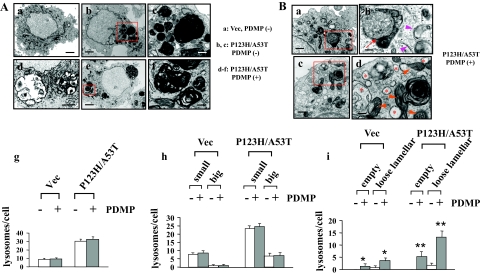
Figure 5.
Ultrastructural analyses of PDMP-treated syn-overexpressing cells. Electron microscopic analyses were performed for P123H β-syn-overexpressing cells transfected with A53T α-syn, followed by treatment with (Ad–Af, Ba–Bd) or without (Ab and Ac) PDMP treatment for 24 hours. Vector-transfected cells were also analyzed as a control (Aa). A: At a low magnification, P123H β-syn-overexpressing cells transfected with A53T α-syn exhibited various sizes of electron-dense inclusion clusters (b and e), whereas fewer lysosomal structures were found in vector-transfected cells (a). c: At a higher magnification, some large electron-dense inclusions were composed of multilamellar myelinosomes whose membranes were tightly packed with each other, whereas other small electron-dense inclusions made a fusion with relatively light dense structures of unknown origins. f: After treatment with PDMP, the tightly packed structures of multilamellar myelinosomes became so loose as to give rise to a gap between each membrane. d: Further, empty inclusion bodies, the contents of which were probably discharged into the cytoplasm, were frequently encountered. Small boxes in b and e are equivalent to c and f, respectively. In g–i, morphometric analysis was performed as described in the Materials and Methods. g: The numbers of lysosomal inclusions were higher in both P123H β-syn-overexpressing cells transfected with A53T α-syn compared with vector-transfected cells. However, there were no significant changes of the numbers of lysosomes before and after PDMP treatment. h: Similar results were obtained when the lysosomal structures were categorized into two groups according to their diameters; small: 400 nm to ∼1 μm, and big: more than 1 μm. In i, the numbers of empty giant inclusion and loose lamellar structures of inclusions were significantly increased after PDMP treatment in P123H β-syn-overexpressing cells transfected with A53T α-syn and to a lesser extent in vector-transfected cells. Data are shown as means ± SD (n = 50 cells). *P < 0.05, **P < 0.01 versus PDMP-untreated cells. B: In PDMP-treated P123H β-syn-overexpressing cells transfected with A53T α-syn, there was extensive formation of autophagic vacuoles (b, arrow) and unclosed double-membrane structures (b, arrowheads). Furthermore, mitochondria (d, arrowheads) and ER (d, asterisks) were frequently swollen and distorted. Small boxes in a and c are equivalent to b and d, respectively. In both A and B, the electron micrograph is representative of at least three independent experiments. Scale bars: 4 μm (Aa, Ab, Ad, Ae, Ba, Bc); 1 μm (Ac, Af, Bb, Bd).

