Figure 1.
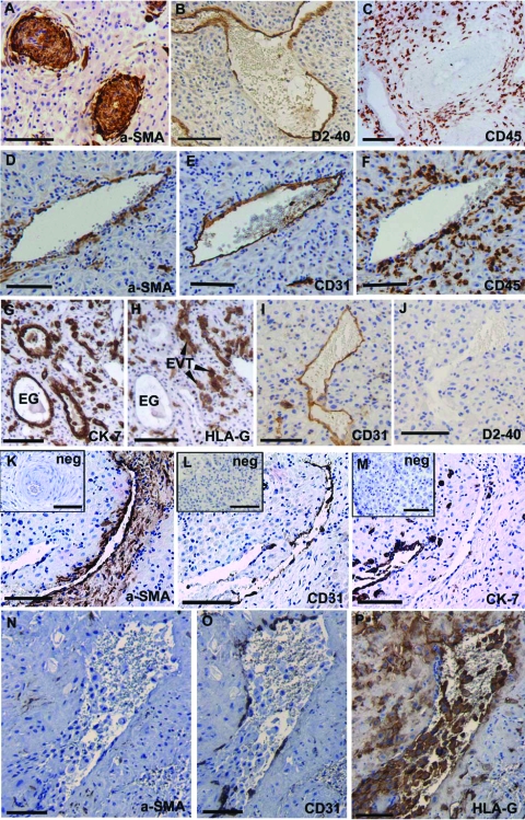
Immunohistochemical identification of remodeling vessels in decidua basalis. A: Unremodeled artery stained for α-smooth muscle actin (SMA). B: Lymphatic vessel stained with D2–40. C: Decidua parietalis stained with CD45 demonstrating the abundance of leukocytes. D–F: Vein exhibiting: (D) a thin layer of VSMC (α-SMA immunostaining), (E) intact endothelium (CD31 immunostaining) and (F) leukocytes clustering around the venous wall (CD45 immunostaining). G: Cytokeratin (CK)−7 immunostaining was present in the glandular epithelium (EG) and extravillous trophoblasts (EVT), while (H) HLA-G immunostaining identified only the EVT. I: CD31 immunostaining stained all endothelial cells and (J) absence of staining for D2–40 on a serial section revealed that this vessel was not lymphatic. K–M: Remodeling vessel exhibiting: (K) disrupted vascular smooth muscle cells (VSMC), (L) loss of endothelium, and (M) absence of EVT in the vessel wall. Inset: Representative negative controls for: (K) α-SMA, (L) CD31, and (M) HLA-G immunohistochemistry. N–P: More extensively remodeled vessel exhibiting: (N) complete loss of VSMC, (O) further loss of endothelium, and (P) EVT presence within the stroma, vessel lumen, and relining the vessel wall. Scale bars: 50 μm (D–F) and 100 μm (A–C and G–P).

