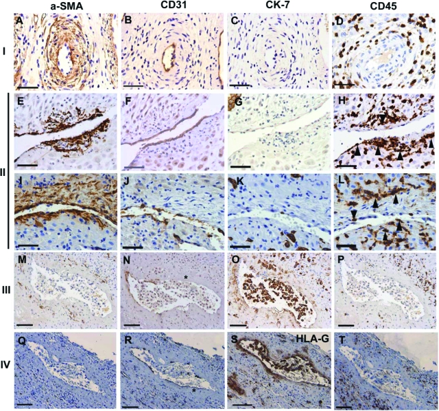
Figure 2.
Stages of vascular remodeling and leukocyte involvement. Remodeling vessels were identified by immunostaining of serial sections with α-smooth muscle actin (SMA), CD31, Cytokeratin (CK)−7/HLA-G and CD45 (leukocyte common antigen). A–D: Stage I: Unremodeled vessels displayed: (A) intact and organized vascular smooth muscle cells (VSMC), (B) intact endothelium, (C) absence of extravillous trophoblast (EVT) from the vessel wall, and (D) leukocytes absent from the vascular wall. E–L: Stage II: Vessels displayed: (E and I) dramatically disrupted and disorganized VSMC, (F and J) loss of endothelium, (G and K) absence of EVT from the vessel wall and lumen and (H and L) leukocyte infiltration into the vascular wall and VSMC layers (arrowheads). M–P: Stage III: Vessels displayed: (M) substantial loss of VSMC, (N) and endothelium, (O) presence of EVT within the vessel lumen and (P) leukocytes were largely absent from the vascular wall. Q–T: Stage IV: Remodeled vessels displayed: (Q) complete loss of VSMC, (R) further (but not complete) loss of endothelium from the vascular wall, (S) EVT relining the vessel and (T) leukocyte absence from the vascular wall. Asterisk denotes fibrinoid. Scale bars; 50 μm (A–L) and 100 μm (M–T).

