Figure 5.
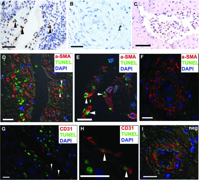
Identification of apoptotic vascular cells during remodeling. TUNEL immunostaining in (A) a stage II remodeling vessel, arrowheads highlight positive nuclei within the vascular wall. No TUNEL positivity was detected in (B) unremodeled or (C) remodeled vessels. D and E: Dual fluorescent staining for α-smooth muscle actin (SMA) and TUNEL, and for (G and H) CD31 and TUNEL revealed that proportion of TUNEL positive cells were also α-SMA positive and CD31 positive (arrowheads denote dual labeled cells). F: No TUNEL positivity was detected in unremodeled vessels. I: Dual stained TUNEL negative control: section was immunostained for α-SMA (red cells) and TUNEL negative control (FITC-labeled dUTPs applied but TdT enzyme omitted). Scale bars: 20 μm (D–I), 50 μm (A and B), and 100 μm (C).

