
Figure 4. SHH-N treatment promotes proliferation in embryonic organotypic retinal explant cultures from orJ mice.
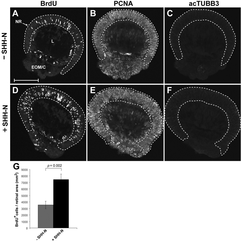
E12.5 explants were cultured in the absence (A–C) or presence (D–F) of SHH-N for 24 hr with BrdU added for the last 30 min. (A, D) BrdU incorporation. (B, E) PCNA immunoreactivity. (C, F) acTUBB3 immunoreactivity. Retinal tissue is contained within the dashed lines. Scale bar: 100 µm. (G) Quantification of BrdU+ cells as a function of retinal area (mm2). Each bar represents the mean ± standard error of the mean (SEM). p values calculated using Student’s paired t-test