
Figure 8. SHH-N treatment promotes proliferation in neonatal explants within 24 hr.
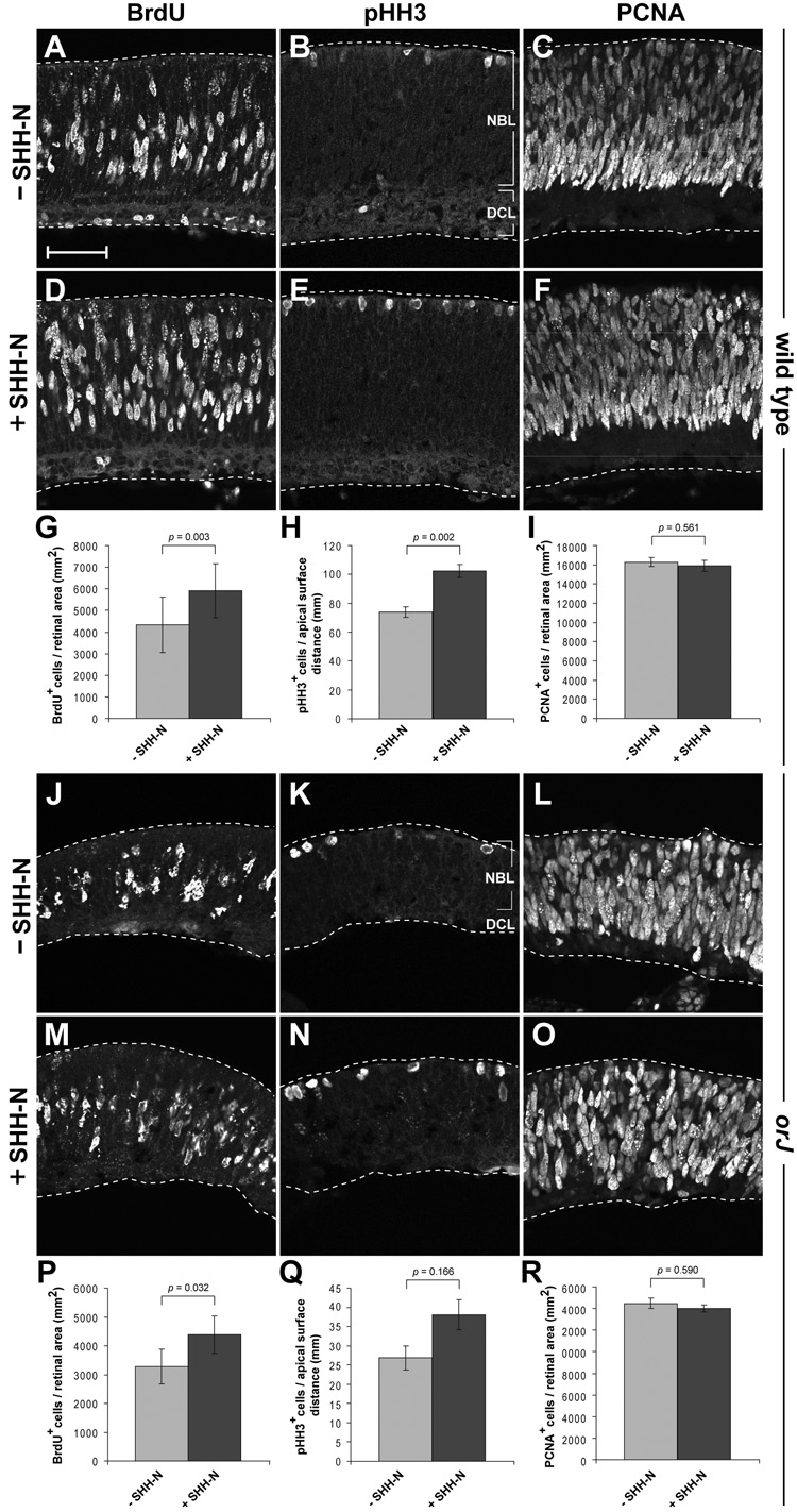
BrdU (A, D), pHH3 (B, E), and PCNA (C, F) immunoreactivity in wild type explants after 24 hr in culture. Quantification of BrdU+ (G), pHH3+ (H), and PCNA+ (I) cells in wild type explants as a function of retinal area (G, I) and length of the apical surface of the retina (H). BrdU (J, M), pHH3 (K, N), and PCNA (L, O) immunoreactivity in orJ explants after 24 hr in culture. Quantification of BrdU+ (P), pHH3+ (Q), and PCNA+ (R) cells in wild type explants as a function of retinal area (P, R) and length of the apical surface of the retina (Q). Bars represent mean ± SEM. p values calculated using Student’s paired t-test. Scale bar: 50 µm.