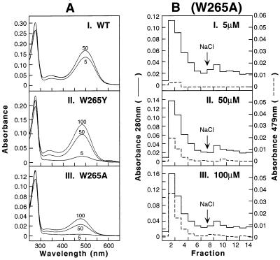
Figure 2.
UV–vis spectra of rhodopsin and rhodopsin mutants formed on retinal treatment of COS-1 cells expressing opsins. Whole-cell suspensions of transfected COS-1 cells were divided and treated with the 11-cis-retinal at concentrations indicated. Pigments were solubilized and purified by immunoaffinity chromatography as described in Methods. (A) The UV–vis absorption spectra are shown, I, WT; II, W265Y, and III, W265A. (B) (W265A), elution profiles from 1D-4 Sepharose column of pigments formed after treatment with 11-cis-retinal at 5 μM (I), 50 μM (II), and 100 μM (III). Shown are the absorbances at 280 nm (solid line) and 479 nm (dotted line) for each fraction. Fractions 1–7 in every case were as eluted by using buffer E (no salt), whereas fractions 8–14 were obtained by using buffer F (140 mM NaCl).

