Fig. 5.
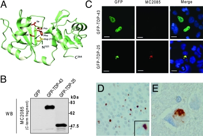
A conformation-dependent TDP-43 antibody that detects C-terminal fragments is specific for pathologic inclusions in human TDP-43 proteinopathies. (A) Three-dimensional modeling of wild-type TDP-43 (amino acids 101–264). The positions of the 8 amino acid residues following the caspase cleavage motif, selected to generate a polyclonal antibody, are indicated. Note that aspartate 219, which is part of the caspase cleavage motif, is buried within the molecule (arrow). (B) Western blot analysis of lysates from HEK293 cells transfected with GFP only, GFP-TDP-43, or GFP-TDP-25 and probed with MC2085. *, Endogenous TDP-43. (C) MC2085 immunostaining of HEK293 cells transfected with GFP-TDP-43 or GFP-TDP-25. Note the absence of staining in cells expressing wild-type TDP-43 compared with those expressing the 25-kDa C-terminal fragment. (Scale bars: 10 μm.) (D and E) Immunostaining of tissue from FTLD-U brain (D) and ALS brain (E) with MC2085 shows cytoplasmic inclusions with little or no nuclear TDP-43. (Magnification: 40×.)

