
Figure 1.
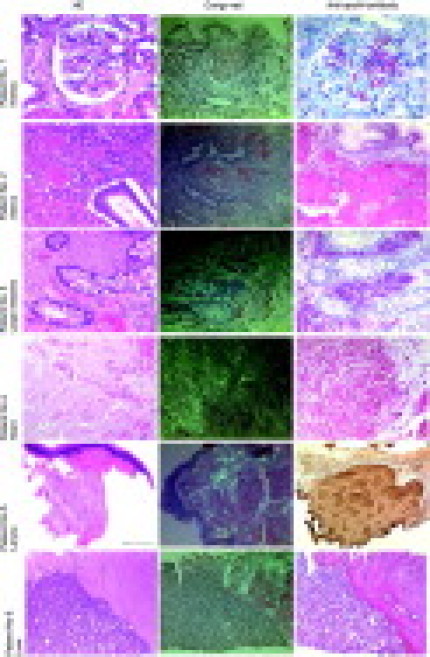
Biopsies from the six patients with AApoAI amyloidosis.Histological examination of the biopsy and resection specimens revealed homogeneous eosinophilic deposits in H&E-stained sections (left panel). Same areas produced a typical apple green birefringence in polarized light after Congo red (CR) staining (center). Staining with an anti-apoAI antibody (right panel) showed a strong and even immunoreactivity of these amyloid deposits in every patient specimen. Sections from the patients 1–4 and 6 were stained with the alkaline phosphatase system (red color), whereas the section from patient No. 5 was stained with a DAB substrate kit (brown color). Scale bar=100 μm.